


Image Integration (Q-BPS™)
The hardware component of IDUS™ includes a module which forms the interface between the sensor array and the ultrasound system. It acquires, conditions, and integrates the signals received by the sensors and performs signal processing for microcalcification acquisition and positioning. The data from the unit is then formatted and co-registered with the traditional ultrasound images, resulting in a 2D B-mode or 3D volume images enhanced by intuitive color coded overlay of spectroscopy (detection and position) information (see sample experimental results below).

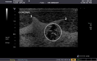
IDUS™ 2D coronal view of the malignant tumor showing detailed structure of line calcification. Points of reference have been marked as “1” and “2”. Images not to scale.
Conventional 2D coronal view of the model malignant tumor performed by a TOSHIBA system, showing the outline of the tumor (see while circle). No details of line calcification was depicted.